
A scan can sometimes find a mass, enlarged lymph node, unusual area of tissue, or another abnormality that raises concern for cancer. But that is not the same as proving that cancer is present. The National Cancer Institute explains that cancer diagnosis often combines imaging with other tests and that a biopsy is often the only way to know for certain.
The same caution works in the other direction. A scan that does not show an obvious tumor is reassuring for the area and problem being evaluated, but it does not universally rule out every cancer. What an imaging test can detect depends on the body part, the size and location of an abnormality, the type of test, and why the study was performed.
Can an Imaging Test Actually Tell You That You Have Cancer?
Imaging can show that something looks abnormal; it cannot always tell exactly what that abnormality is. A lesion may be benign, inflammatory, infectious, traumatic, or cancerous. The American Cancer Society notes that imaging tests can find masses or suspicious changes but sometimes cannot show for sure whether a change is caused by cancer.
That distinction matters when a report uses words such as “mass,” “lesion,” “nodule,” “indeterminate,” or “suspicious.” Those terms describe an imaging finding and its level of concern. They are not interchangeable with a pathology-confirmed cancer diagnosis.
Imaging also has a detection threshold. Very small collections of cancer cells may be below what a scan can display, and some tumors are harder to see because of their location or the surrounding anatomy. A normal result should therefore be interpreted in the context of the specific clinical question rather than as a universal cancer clearance.
What a CT Scan Can Show When Cancer Is a Concern
CT is the strongest of these three modalities for many broad internal evaluations because it creates detailed cross-sectional images of organs, bones, soft tissues, and blood vessels. The National Cancer Institute describes CT as a tool that can help identify tumors, show their location, contribute to staging, and guide biopsy procedures.
Depending on the part of the body being scanned, CT may reveal a suspicious mass, enlarged lymph nodes, an organ abnormality, unexpected fluid, obstruction, bleeding, or another complication. Contrast material is sometimes used because it can make certain tissues and boundaries easier to distinguish.
Can a CT scan detect cancer?
Yes, CT can detect many abnormalities that may represent cancer. The American Cancer Society explains that CT can show a tumor’s shape, size, location, and relationship to nearby structures. But the answer is not “CT detects every cancer.” A small tumor, an abnormality outside the scanned region, or a cancer that does not create a clearly visible structural change may not be apparent on that particular exam.
The reason for the scan matters too. An emergency CT ordered for severe abdominal pain is designed and interpreted around the urgent problem being evaluated. It can reveal an unexpected mass, but it is not the same as a comprehensive cancer-screening examination.
Why a CT finding may still need more testing
A radiologist may be able to say that a finding is clearly benign, but other findings remain indeterminate. When an abnormality cannot be fully characterized, the report may recommend comparison with prior scans, targeted follow-up imaging, specialist review, or tissue sampling. RadiologyInfo shows how CT reports may recommend additional imaging or biopsy when a lesion remains uncertain.
That follow-up is not a contradiction of the CT result. It is how imaging is supposed to work: first identify and describe the abnormality, then use the most appropriate next test to determine what it represents.
Can an X-Ray Show Cancer or a Tumor?
Sometimes. A plain X-ray can reveal certain tumors or changes caused by cancer, particularly in the lungs or bones, but it provides less anatomical detail than CT. The American Cancer Society explains that standard X-rays can help identify problems including some cancers, while CT often gives better pictures of soft tissues.
An X-ray may therefore raise suspicion rather than settle the diagnosis. A chest X-ray, for example, might show a lung opacity or mass that needs CT evaluation. It can also miss small or obscured abnormalities. RadiologyInfo specifically notes that small cancers may not appear on a conventional chest X-ray.
So if symptoms, examination findings, or another test keep concern high, a normal routine X-ray does not necessarily end the evaluation.
Can Ultrasound Detect Cancer?
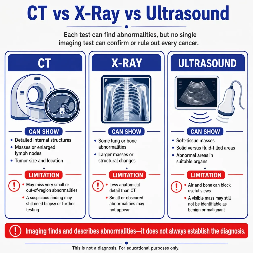
Ultrasound can detect and characterize many abnormalities in parts of the body that are well suited to sound-wave imaging. It can show soft-tissue structures, fluid collections, cystic versus solid features, and masses in certain organs. The American Cancer Society notes that ultrasound can help find tumors, distinguish some fluid-filled cysts from solid masses, and guide biopsy needles.
Its limitations are just as important. Ultrasound is not equally effective in every body region; air and bone interfere with sound waves, and some deep structures are difficult to evaluate. Even when ultrasound shows a mass, the image may not be able to determine whether it is benign or malignant.
In practice, ultrasound often helps answer a focused question: Is there a mass? Is it solid or fluid-filled? Is there an abnormal area that needs a different imaging test or tissue sampling? It is not a universal “cancer yes-or-no” test.
What Happens When Imaging Finds Something Suspicious?
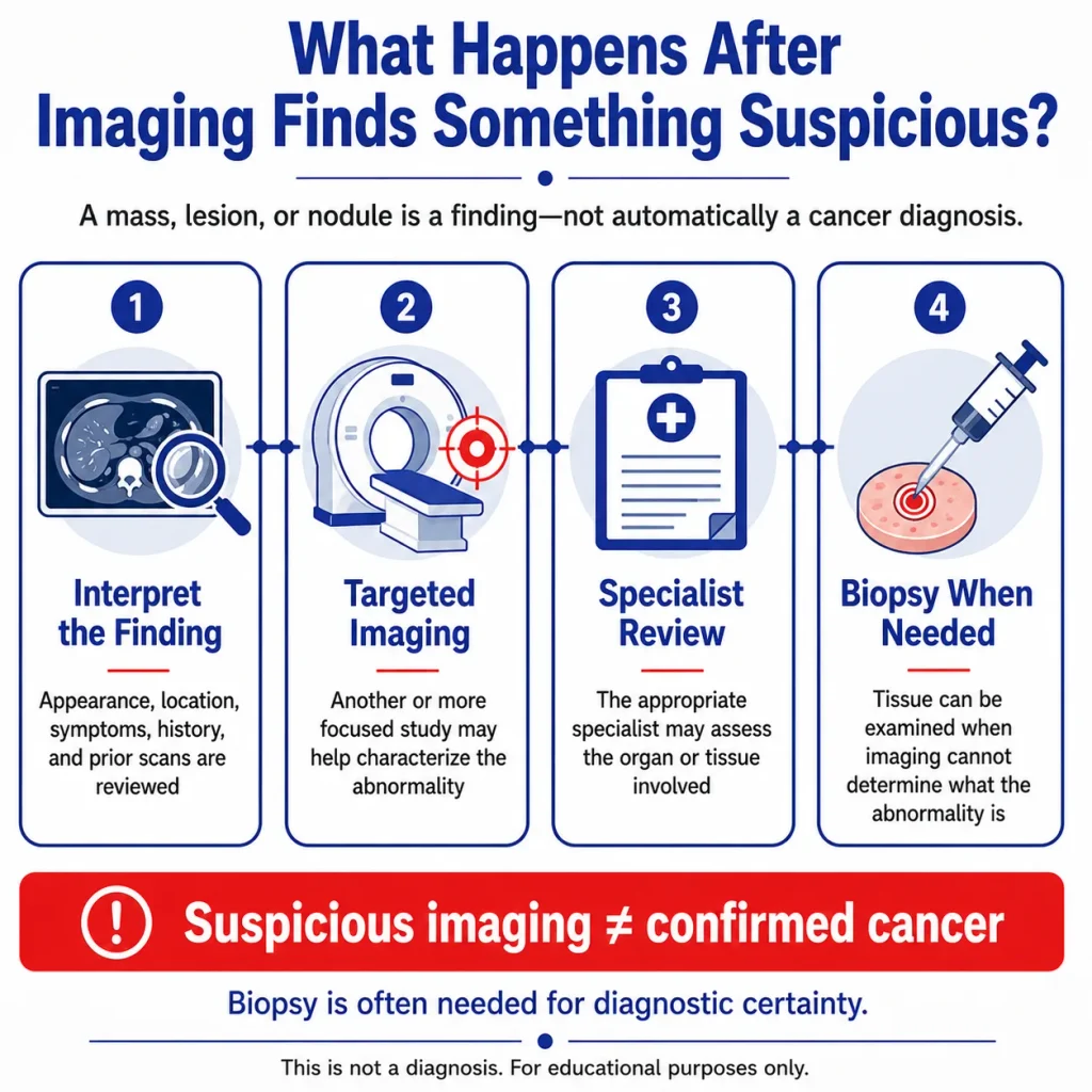
If a report says a mass or lesion is suspicious, the next step is to determine how much certainty the imaging provides and what information is still missing. “Suspicious” and “confirmed cancer” are different stages of evaluation.
1. The finding is interpreted in context. The radiologist and treating clinician consider the appearance, location, symptoms, medical history, prior imaging, and any relevant lab results.
2. Additional imaging may be recommended. A more targeted study may be needed to characterize the abnormality or check another body region. The exact test depends on what was found; not every patient needs another scan.
3. Specialist assessment may be needed. The appropriate specialist depends on the organ or tissue involved and the level of concern.
4. A biopsy may be necessary. When imaging cannot confidently determine what a suspicious area is, tissue may be sampled for laboratory examination. RadiologyInfo notes that biopsy is commonly used when imaging cannot clearly define whether an abnormality is benign or malignant.
Biopsy guidance from RadiologyInfo explains that CT or ultrasound can also be used to guide a needle precisely to the abnormal area.
The workup is also disease-specific. For example, plasma-cell cancers may require blood and urine testing in addition to imaging; Post Oak ER covers those details separately in its guide to multiple myeloma blood and urine tests. Keeping those roles separate prevents any single test from being given more certainty than it actually provides.
Emergency Imaging and a Cancer Workup Are Not the Same Thing
CT, X-ray, or ultrasound performed during an emergency visit is generally chosen to evaluate the acute problem that brought the patient in: severe pain, trouble breathing, bleeding, neurologic symptoms, injury, or another urgent change. The immediate question is what needs attention now.
That scan can still reveal a mass or other concerning finding incidentally. If it does, the emergency team can address urgent complications and explain what follow-up is needed. But an ER visit is not a substitute for a complete cancer-screening, biopsy, staging, or oncology pathway.
For Houston-area patients, Post Oak ER provides 24/7 emergency care and offers on-site CT, X-ray, and ultrasound as part of emergency evaluations when clinically appropriate. If emergency imaging identifies a suspicious abnormality, the immediate medical issue can be evaluated and appropriate follow-up recommended, while definitive cancer diagnosis may continue after the acute visit.
Frequently Asked Questions
Can a CT scan detect cancer anywhere in the body?
CT can evaluate many body regions and can detect numerous tumors, but no single CT scan examines every possible site with equal sensitivity. What it can show depends on the scanned area, the tumor’s size and location, the protocol used, and the clinical question.
Can you have cancer even if a CT scan is normal?
Yes. A normal CT means no concerning abnormality was identified within the limits of that examination; it does not rule out every cancer in every location. If symptoms or other findings remain concerning, a clinician may recommend a different or more targeted evaluation.
Can an X-ray show a cancerous tumor?
It can show some tumors or cancer-related changes, particularly in the lungs or bones. However, routine X-rays provide less detail than CT, and smaller or obscured lesions may not be visible.
Can ultrasound tell whether a mass is cancerous?
Sometimes ultrasound features can make a mass look more or less concerning, but ultrasound cannot reliably label every mass as benign or malignant. Additional imaging, specialist evaluation, or biopsy may be needed.
If imaging finds a mass, does that mean it is cancer?
No. A mass is an abnormal finding, not automatically a cancer diagnosis. Benign growths, cysts, inflammation, infection, and other conditions can also produce masses or mass-like appearances.
Why might a biopsy be needed after an abnormal CT, X-ray, or ultrasound?
A biopsy allows tissue from the suspicious area to be examined in the laboratory. It is often used when imaging can locate and describe an abnormality but cannot determine with enough certainty what the abnormal tissue actually is.